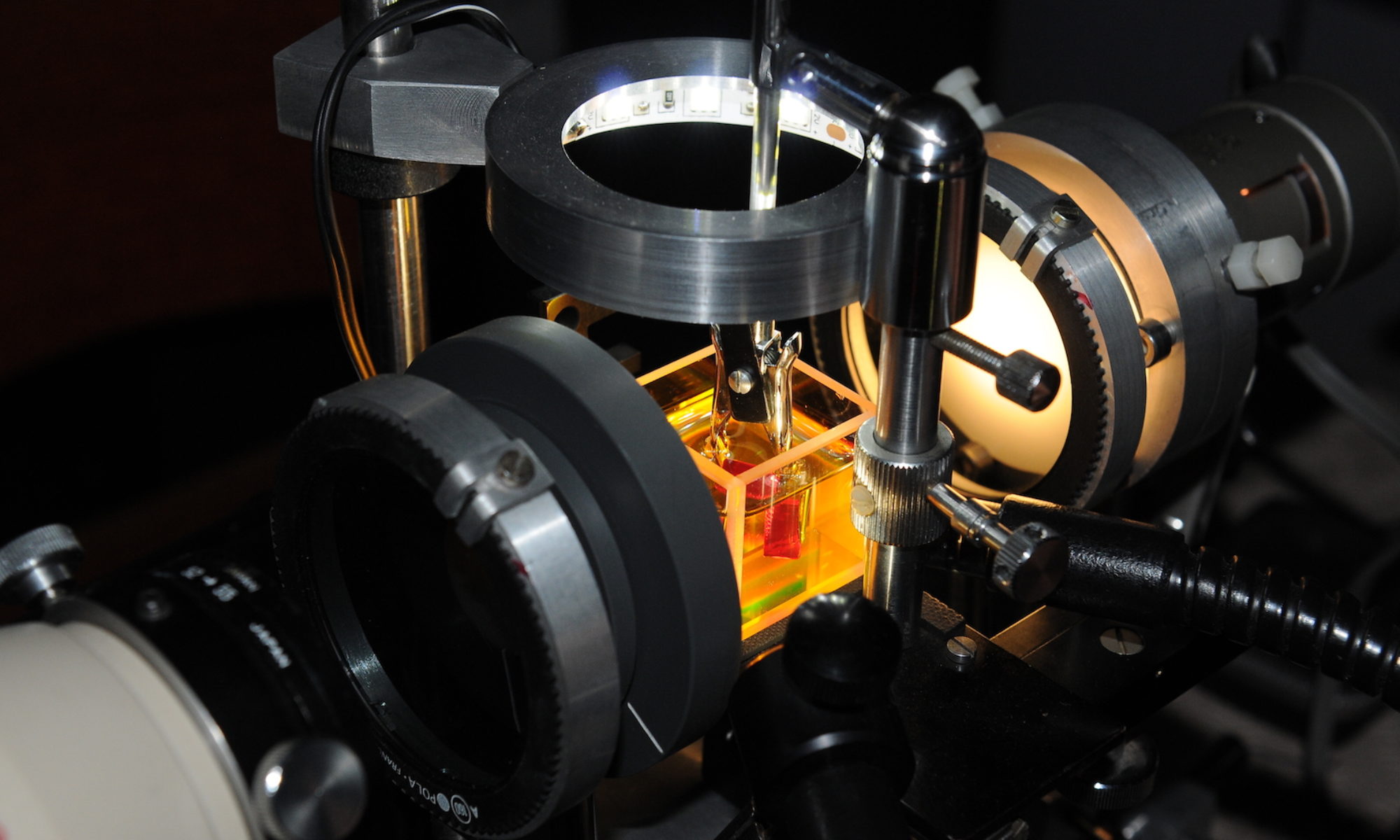
This image shows a Lily Pad inclusion in peridot. One can notice that only the inclusion itself can be seen. The Lily Pad was observed on microscope without the use of immersion cell. Two very small fiber optic lights were used to only highlight the inclusion by it self. By using this technique one can avoid the hazard of internal reflections. The image was captured with a digital ocular of Kern & Sohn, which allows working in very low light conditions.

