Welcome to Case Studies
QUESTION OF THE WEEK
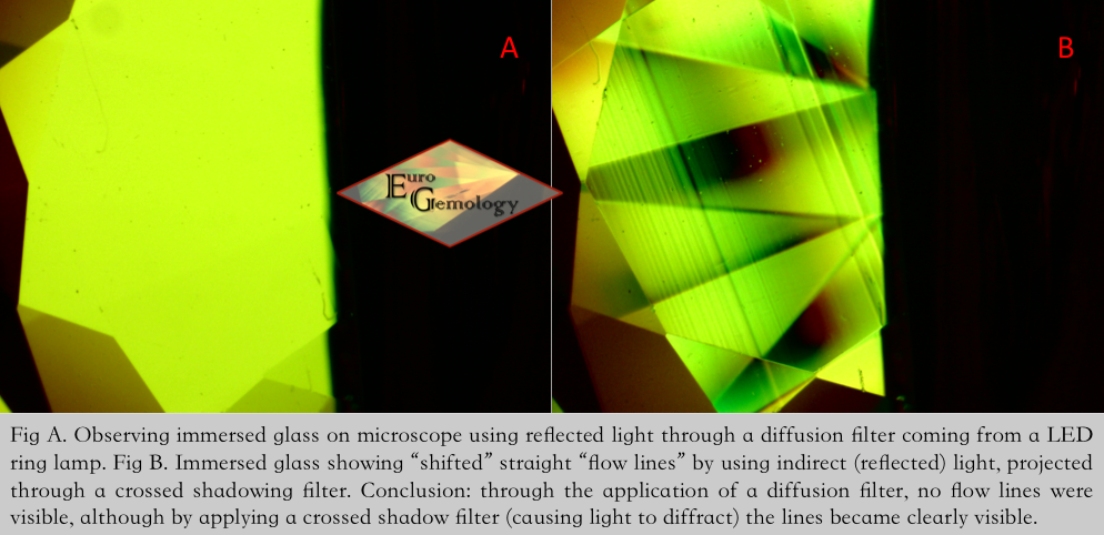
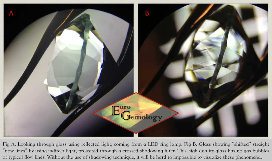
WHAT IS THIS?

CATEGORY: Mounted Gemstones
LEVEL: Medium to Difficult
PURPOSE: Recycling
DESCRIPTION
This ring was sold by an auction house. The gemstone mounted in the ring was described as being a “colored center stone” mounted on an 18 karat golden ring. The item was bought to be recycled.
OBSERVATIONS
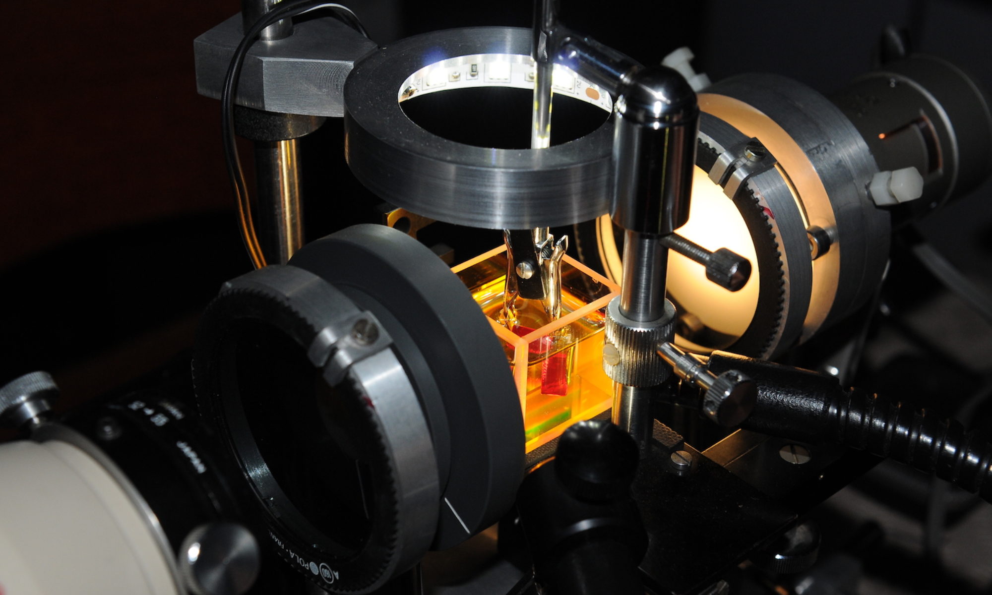
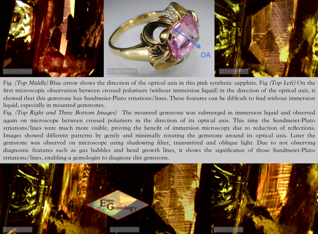
This pink transparent center “gemstone” mounted on a ring doesn’t show any external abnormalities. It is a facetted gemstone. Through examination in immersion polariscope with ¼ lambda retardation plate, it visually reveals the gemstone being uniaxial. In normal cases, it’s fairly easy to determine the optic sign with the retardation plate, but in this case due to the surrounding alloy, it is not visible. Therefore, the optic sign has to be determined through refractometer readings. By usage of the gemological refractometer, results show to have following readings: ne = 1,760 and no= 1,768 with a birefringence of 0,008. As the HIGH RI is constant, it proves that the gemstone is uniaxial (-1). By observing the gemstone through dichroscope, a mild dichroism can be noticed by manner of the colors being yellow and pink. Under UVL, the center gemstone shows a strong red fluorescence and a strong blue fluorescence under UVS.
It was not possible to verify the specific gravity/density since this gemstone is mounted in a ring. Both side stones tested negative with the diamond tester. No further research on them was requested.
Gold was tested with a conventional acid test and shows to be 18 karat.
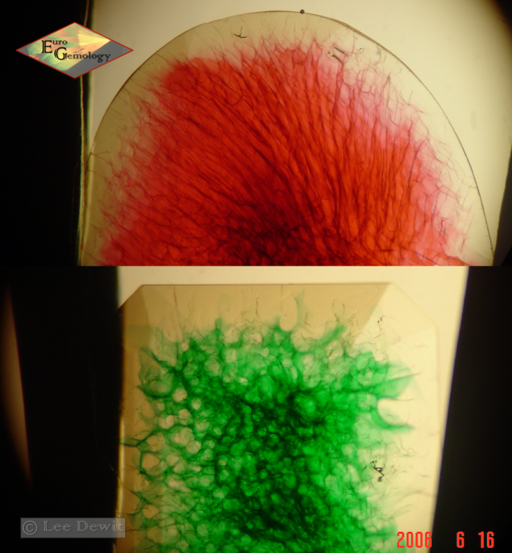
During the first microscopic observation between crossed polarisers (without immersion liquid) in the direction of the optical axis, it showed that this stone has Sandmeier-Plato striations/lines.
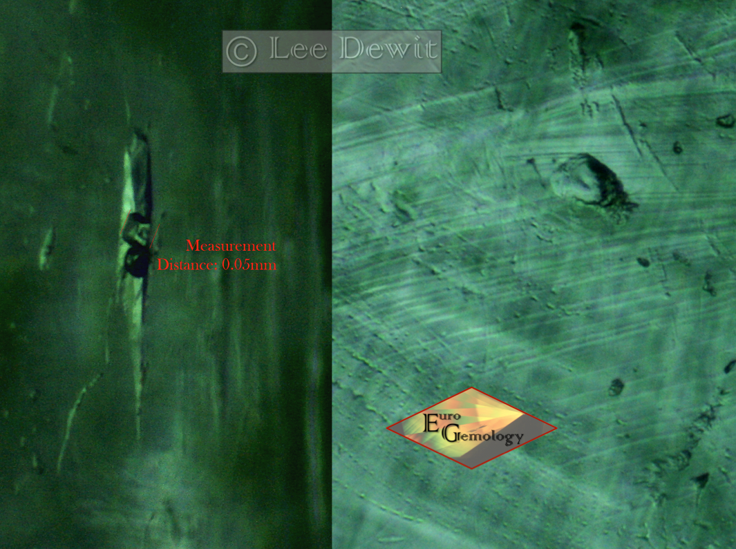
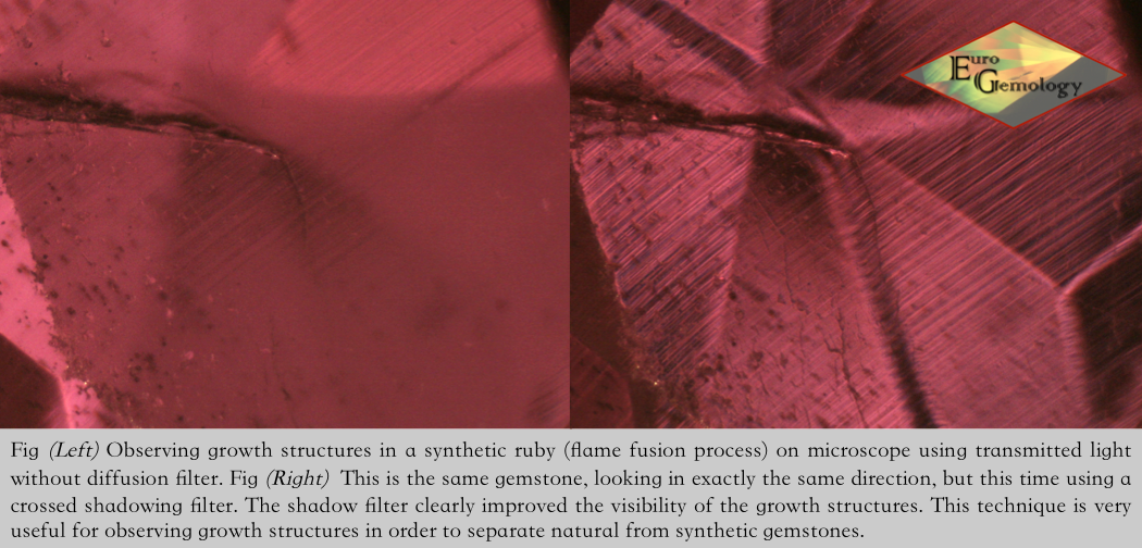
Secondly, the mounted gemstone was submerged in immersion liquid and observed again on microscope using, shadowing filter, transmitted and oblique light. It revealed that this gemstone has no gas bubbles and no sign of bend growth lines.
RESULT
It is an 18 karat golden ring containing two diamond imitations with a central gemstone being a pink synthetic sapphire.
CONCLUSION
It is rather difficult to observe Sandmeier-Plato striations/lines in gemstones mounted in jewelry without the use of immersion liquid. In this case they were clearly visible without the use of immersion liquid. Observing them can be very important in light colored corundum (e.g. colorless, light yellow and light pink sapphires) to separate them from their synthetic counterparts. In synthetic ruby and blue sapphires made through flame fusion, gas bubbles and bend growth lines are to be expected. When those features are absent in very clear stones, Sandmeier-Plato striations/lines would be very indicative dealing with synthetic corundum made through flame fusion procedure. When a strong UVS (blue) and UVL (red) fluorescence is present the Sandmeier-Plato striations/lines and fluorescence would both become diagnostic features.